
What is the Acid-Fast Stain, and how do we use it?
Do you enjoy working in a science lab, and are you interested in helping diagnose patients? If you choose an exciting job in a medical diagnostics lab, the Acid-Fast, or Ziehl-Neelsen stain, is a bacterial staining technique that you may be called to perform often! If you choose another amazing career, this is still very useful information.
The purpose of the stain is to determine whether or not the bacteria’s cell wall has mycolic acids. Mycolic acids are long fatty acid chains with 60 to 90 carbon atoms. They are present in species of the genera Nocardia and Mycobacterium. Nocardia may cause Nocardiosis, a dangerous infection that starts in the lungs and may spread to other organs, such as the brain, kidneys, or heart. Mycobacterium tuberculosis is the bacteria responsible for - you guessed it - tuberculosis(TB), an infectious lung disease that may also spread to other body parts.
Let’s say a patient comes in with symptoms such as fever, night sweats, and a long-lasting cough. It might be a good idea to do a test to rule out Nocardiosis or TB! Now how would we accomplish this? The patient would give us a sample - typically blood, urine, stool, or sputum (mucus they coughed up, doesn’t that sound fun)! Then, you would carefully do the lab work - spread the sample on a plate and allow the bacteria to multiply for about two days. After that time, place some bacteria on a microscope slide and stain it.
You start by applying the primary stain: carbol fuchsin, which is pink. Next, you decolorize with acid alcohol. Finally, you apply the counterstain, something with a blue or purple color. One commonly used counterstain is methylene blue.
If the bacteria is acid-fast, it will appear pink. Non-acid fast bacteria will be purple or blue in color, depending on which counterstain you used. A positive result means it has mycolic acid, which helps the cell wall resist acid alcohol and retain the pink color. If negative, it lacks mycolic acid and allows the acid alcohol to wash out the carbol fuchsin, so the counterstain can come in and make the cells blue or purple!
If the bacterial cells are pink, the patient likely has TB. If they’re only partially pink, they probably have Nocardiosis. If the cells are blue or purple, something else may be making our patient sick. If done correctly, this stain is a great step in answering questions and getting to the root of the problem!
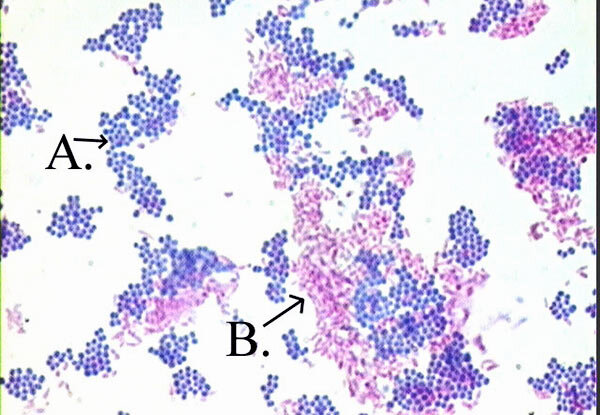
A shows non-acid-fast bacteria (purple). B shows acid-fast bacteria (pink). They are differentiated by the color produced by the Acid-fast stain.
Picture Source: microbiologyinfo.com