
What Are Capsules? How Do We Stain Them?
While bacteria are tiny creatures, they have developed some creative defenses when they find themselves in tough conditions! Let's suppose they are facing dehydration in a very dry environment, or they are being attacked by immune cells while they are causing an infection in a host (which isn't so nice of the bacteria, is it?). Neither of these situations sound like fun. So how could the bacteria survive? One possible solution is to form capsules! Capsules are slippery coatings around the bacterium, typically made of polysaccharides(many sugars bound together). Many capsules are hydrophilic(water-loving) and retain water for the cell. They may also help the bacteria escape phagocytosis, or attacks by the immune system. To put it simply, they mask the bacteria so the immune system doesn't recognize it as a threat.
In the microbiology lab, if we want to check whether a species of bacteria produces capsules, we can perform the capsule staining procedure. There are different choices of stains we can use for this procedure, I'll just discuss one pair. In any case, one stain would be positively charged, and the other negatively charged.
Start with two microscope slides. Place the bacteria on one end of one slide, then emulsify(mix) with one or two drops of Congo Red stain. Use the second slide to spread the mixture across the first slide. Once it air-dries, apply Maneval's capsule stain to the slide for one minute, and rinse it off with distilled water. Under the microscope, our slide and our bacteria will appear as a darker color. Capsules will appear as transparent ovals around the bacteria.
Remember this from chemistry? Similar charges repel each other and opposite charges attract! The Congo Red is acidic/negatively charged, so it is repelled from the negatively charged bacteria. In contrast, the Maneval's stain is basic/positively charged and binds to the cell. Capsules have a neutral net charge, so they will not bind either of the stains.
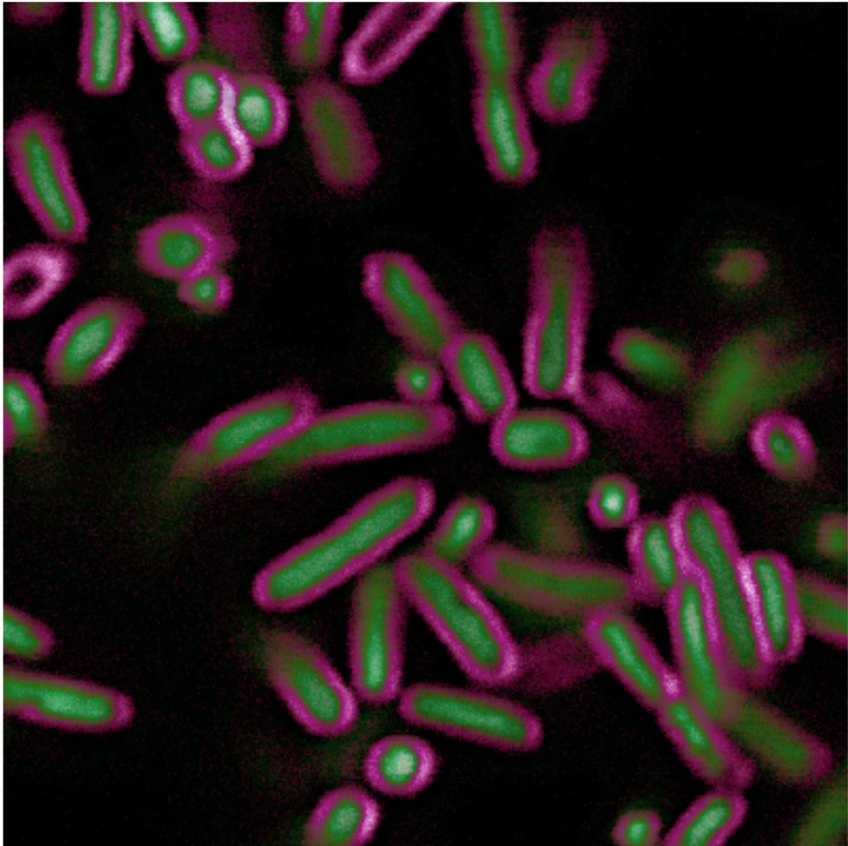
E. coli stained with BactoView™ Live Green (stains DNA) and SynaptoRed™ (stains the membrane)
Picture Source: biotium.com